What is a Corona Virus and do they commonly cause influenza?
by
Robert Gorter, MD, PhD, et al.
Coronaviruses are a group of related RNA viruses that cause diseases in mammals and birds. In humans, these viruses cause respiratory tract infections that can range from mild to lethal. Mild illnesses include some cases of the common cold (which is also caused by other viruses, predominantly rhinoviruses), while more lethal varieties can cause SARS, MERS, and COVID-19. Symptoms in other species vary: in chicken, they cause an upper respiratory tract disease, while in cows and pigs they cause diarrhea.
Although know for almost 100 years and after multiple attempts, there are as of yet no vaccines or antiviral drugs to prevent or treat neither human coronavirus infections nor HIV.
Coronaviruses constitute the subfamily Orthocoronavirinae, in the family Coronaviridae, order Nidovirales, and realm Riboviria.[5][6] They have enveloped viruses with a positive-sense single-stranded RNA genome and a nucleocapsid of helical symmetry.[7] The genome size of coronaviruses ranges from approximately 26 to 32 kilobases, one of the largest among RNA viruses.[8] They have characteristic club-shaped spikes that project from their surface, which in electron micrographs create an image reminiscent of the solar corona, from which their name derives.[9]
Corona Virus is an RNA virus that replicates in the cytoplasm of the host cell. HIV, for instance, is an RNA Retrovirus and builds itself into the chromosomes (see the second part of this summary).
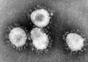
Coronaviruses are a group of viruses that have a halo, or crown-like (corona) appearance when viewed under an electron microscope. The coronavirus is now recognized as the etiologic agent of the 2003 SARS outbreak. Additional specimens are being tested to learn more about this coronavirus, and its etiologic link with Severe Acute Respiratory Syndrome.
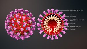
Cross-sectional model of a coronavirus
Etymology
The name “coronavirus” is derived from Latin corona, meaning “crown” or “wreath”, itself a borrowing from Greek κορώνη korṓnē, “garland, wreath”.[10][11] The name was coined by June Almeida and David Tyrrell who first observed and studied human coronaviruses.[12] The word was first used in print in 1968 by an informal group of virologists in the journal Nature to designate the new family of viruses.[9] The name refers to the characteristic appearance of virions (the infective form of the virus) by electron microscopy, which has a fringe of large, bulbous surface projections creating an image reminiscent of the solar corona or halo.[9][12] This morphology is created by the viral spike peplomers, which are proteins on the surface of the virus.[13]
History
Coronaviruses were first discovered in the 1930s when an acute respiratory infection of domesticated chickens was shown to be caused by the infectious bronchitis virus (IBV).[14] Arthur Schalk and M.C. Hawn described in 1931 a new respiratory infection of chickens in North Dakota. The infection of new-born chicks was characterized by gasping and listlessness. The chicks’ mortality rate was 40–90%.[15] Fred Beaudette and Charles Hudson six years later successfully isolated and cultivated the infectious bronchitis virus which caused the disease.[16] In the 1940s, two more animal coronaviruses, mouse hepatitis virus (MHV) and transmissible gastroenteritis virus (TGEV), were isolated.[17] It was not realized at the time that these three different viruses were related.[18]
Human coronaviruses were discovered in the early 1960s.[19][20] They were isolated using two different methods in the United Kingdom and the United States.[21] E.C. Kendall, Malcolm Byone, and David Tyrrell working at the Common Cold Unit of the British Medical Research Council in 1960 isolated from a boy a novel common cold virus B814.[22][23][24] The virus was not able to be cultivated using standard techniques that had successfully cultivated rhinoviruses, adenoviruses, and other known common cold viruses. In 1965, Tyrrell and Byone successfully cultivated the novel virus by serially passing it through the organ culture of the human embryonic trachea.[25] The new cultivating method was introduced to the lab by Bertil Hoorn.[26] The isolated virus when intranasally inoculated into volunteers caused a cold and was inactivated by ether which indicated it had a lipid envelope.[22][27] Around the same time, Dorothy Hamre[28] and John Procknow at the University of Chicago isolated a novel cold virus 229E from medical students, which they grew in kidney tissue culture. The novel virus 229E, like the virus strain B814, when inoculated into volunteers caused a cold and was inactivated by ether.[29]
The two novel strains B814 and 229E were subsequently imaged by electron microscopy in 1967 by Scottish virologist June Almeida at St. Thomas Hospital in London.[30][31] Almeida through electron microscopy was able to show that B814 and 229E were morphologically related by their distinctive club-like spikes. Not only were they related to each other, but they were morphologically related to the infectious bronchitis virus (IBV).[32] A research group at the National Institute of Health the same year was able to isolate another member of this new group of viruses using organ culture and named the virus strain OC43 (OC for organ culture).[33] Like B814, 229E, and IBV, the novel cold virus OC43 had distinctive club-like spikes when observed with the electron microscope.[34][35]
The IBV-like novel cold viruses were soon shown to be also morphologically related to the mouse hepatitis virus.[17] This new group of IBV-like viruses came to be known as coronaviruses after their distinctive morphological appearance.[9] Human coronavirus 229E and human coronavirus OC43 continued to be studied in subsequent decades.[36][37] The coronavirus strain B814 was lost. It is not known which present human coronavirus it was.[38] Other human coronaviruses have since been identified, including SARS-CoV in 2003, HCV NL63 in 2004, HCV HKU1 in 2005, MERS-CoV in 2012, and SARS-CoV-2 in 2019.[39][40] There have also been a large number of animal coronaviruses identified since the early 1960s.[5]
The life cycle of a coronavirus
Infection begins when the viral spike protein attaches to its complementary host cell receptor. After attachment, a protease of the host cell cleaves and activates the receptor-attached spike protein. Depending on the host cell protease available, cleavage and activation allow the virus to enter the host cell by endocytosis or direct fusion of the viral envelope with the host membrane.[50]
Genome translation
On entry into the host cell, the virus particle is uncoated, and its genome enters the cell cytoplasm. The coronavirus RNA genome has a 5′ methylated cap and a 3′ polyadenylated tail, which allows it to act as a messenger RNA and be directly translated by the host cell’s ribosomes. The host ribosomes translate the initial overlapping open reading frames ORF1a and ORF1b of the virus genome into two large overlapping polyproteins, pp1a and pp1ab.[43]
SARS-CoV genome and proteins
The larger polyprotein pp1ab is a result of a -1 ribosomal frameshift caused by a slippery sequence (UUUAAAC) and a downstream RNA pseudoknot at the end of open reading frame ORF1a.[51] The ribosomal frameshift allows for the continuous translation of ORF1a followed by ORF1b.[43]
The polyproteins have their proteases, PLpro and 3CLpro, which cleave the polyproteins at different specific sites. The cleavage of polyprotein pp1ab yields 16 nonstructural proteins (nsp1 to nsp16). Product proteins include various replication proteins such as RNA-dependent RNA polymerase (nsp12), RNA helicase (nsp13), and exoribonuclease (nsp14).[43]
Replicase-transcriptase
A number of the nonstructural proteins coalesce to form a multi-protein replicase-transcriptase complex. The main replicase-transcriptase protein is the RNA-dependent RNA polymerase (RdRp). It is directly involved in the replication and transcription of RNA from an RNA strand. The other nonstructural proteins in the complex assist in the replication and transcription process. The exoribonuclease nonstructural protein, for instance, provides extra fidelity to replication by providing a proofreading function which the RNA-dependent RNA polymerase lacks.[52]
Replication – One of the main functions of the complex is to replicate the viral genome. RdRp directly mediates the synthesis of negative-sense genomic RNA from the positive-sense genomic RNA. This is followed by the replication of positive-sense genomic RNA from the negative-sense genomic RNA.[43]
Transcription – The other important function of the complex is to transcribe the viral genome. RdRp directly mediates the synthesis of negative-sense subgenomic RNA molecules from the positive-sense genomic RNA. This process is followed by the transcription of these negative-sense subgenomic RNA molecules to their corresponding positive-sense mRNAs.[43] The subgenomic mRNAs form a “nested set” which have a common 5′-head and partially duplicate 3′-end.[53]
Recombination – The replicase-transcriptase complex is also capable of genetic recombination when at least two viral genomes are present in the same infected cell.[53] RNA recombination appears to be a major driving force in determining genetic variability within a coronavirus species, the capability of a coronavirus species to jump from one host to another and, infrequently, in determining the emergence of novel coronaviruses.[54] The exact mechanism of recombination in coronaviruses is unclear but likely involves template switching during genome replication.[54]
Assembly and release
The replicated positive-sense genomic RNA becomes the genome of the progeny viruses. The mRNAs are gene transcripts of the last third of the virus genome after the initial overlapping reading frame. These mRNAs are translated by the host’s ribosomes into the structural proteins and several accessory proteins.[43] RNA translation occurs inside the endoplasmic reticulum. The viral structural proteins S, E, and M move along the secretory pathway into the Golgi intermediate compartment. There, the M proteins direct most protein-protein interactions required for the assembly of viruses following its binding to the nucleocapsid. Progeny viruses are then released from the host cell by exocytosis through secretory vesicles. Once released the viruses can infect other host cells.[55]
Transmission
Infected carriers can shed viruses into the environment. The interaction of the coronavirus spike protein with its complementary cell receptor is central in determining the tissue tropism, infectivity, and species range of the released virus.[56][57] Coronaviruses mainly target epithelial cells.[5] They are transmitted from one host to another host, depending on the coronavirus species, by either an aerosol, fomite or fecal-oral route.[58]
Human coronaviruses infect the epithelial cells of the respiratory tract, while animal coronaviruses generally infect the epithelial cells of the digestive tract.[5] SARS coronavirus, for example, infects via an aerosol route,[59] the human epithelial cells of the lungs by binding to the angiotensin-converting enzyme 2 (ACE2) receptor.[60] Transmissible gastroenteritis coronavirus (TGEV) infects, via a fecal-oral route,[58] the pig epithelial cells of the digestive tract by binding to the alanine aminopeptidase (APN) receptor.[43]
Origins of human coronaviruses with possible intermediate hosts
The most recent common ancestor (MRCA) of all coronaviruses is estimated to have existed as recently as 8,000 BCE, although some models place the common ancestor as far back as 55 million years or more, implying long term coevolution with bat and avian species.[63] The most recent common ancestor of the alphacoronavirus line has been placed at about 2,400 BCE, of the betacoronavirus line at 3,300 BCE, of the gammacoronavirus line at 2,800 BCE, and of the delta coronavirus line at about 3,000 BCE. Bats and birds, as warm-blooded flying vertebrates, are an ideal natural reservoir for the coronavirus gene pool (with bats the reservoir for alphacoronaviruses and betacoronavirus – and birds the reservoir for gammacoronaviruses and deltacoronaviruses). The large number and global range of bat and avian species that host viruses have enabled extensive evolution and dissemination of coronaviruses.[64]
Many human coronaviruses have their origin in bats.[65] The human coronavirus NL63 shared a common ancestor with a bat coronavirus (ARCoV.2) between 1190 and 1449 CE.[66] The human coronavirus 229E shared a common ancestor with a bat coronavirus (GhanaGrp1 Bt CoV) between 1686 and 1800 CE.[67] More recently, alpaca coronavirus and human coronavirus 229E diverged sometime before 1960.[68] MERS-CoV emerged in humans from bats through the intermediate host of camels.[69] MERS-CoV, although related to several bat coronavirus species, appears to have diverged from these several centuries ago.[70] The most closely related bat coronavirus and SARS-CoV diverged in 1986.[71] A possible path of evolution of SARS coronavirus and keen bat coronaviruses is that SARS-related coronaviruses coevolved in bats for a long time. The ancestors of SARS-CoV first infected leaf-nose bats of the genus Hipposideridae; subsequently, they spread to horseshoe bats in the species Rhinolophidae, then to Asian palm civets, and finally to humans.[72][73]
Unlike other beta coronaviruses, bovine coronavirus of the species Betacoronavirus 1 and subgenus Embecovirus is thought to have originated in rodents and not in bats.[65][74] In the 1790s, equine coronavirus diverged from the bovine coronavirus after a cross-species jump.[75] Later in the 1890s, human coronavirus OC43 diverged from bovine coronavirus after another cross-species spillover event.[76][75] It is speculated that the flu pandemic of 1890 may have been caused by this spillover event, and not by the influenza virus, because of the related timing, neurological symptoms, and unknown causative agent of the pandemic.[77] Besides causing respiratory infections, human coronavirus OC43 is also suspected of playing a role in neurological diseases.[78] In the 1950s, the human coronavirus OC43 began to diverge into its present genotypes.[79] Phylogenetically, the mouse hepatitis virus (Murine coronavirus), which infects the mouse’s liver and central nervous system,[80] is related to human coronavirus OC43 and bovine coronavirus. Human coronavirus HKU1, like the aforementioned viruses, also has its origins in rodents.[65]
Infection in humans

Seasonal distribution of HCV-NL63 and other Coronaviruses in Germany shows a preferential detection from November to March; extremely similar in the case of COVID-19
Common cold
The common cold, also known simply as a cold, is a viral infectious disease of the upper respiratory tract that primarily affects the nose. The throat, sinuses, and larynx may also be affected. Signs and symptoms may appear less than two days after exposure to the virus. These may include coughing, sore throat, runny nose, sneezing, headache, and fever. People usually recover in seven to ten days, but some symptoms may last up to three weeks. Occasionally, those with other health problems may develop pneumonia.
Well over 200 virus strains are implicated in causing the common cold, with rhinoviruses being the most common. They spread through the air (aerosols) during close contact with infected people or indirectly through contact with objects in the environment, followed by transfer to the mouth or nose. Risk factors include going to child care facilities, not sleeping well, and psychological stress. The symptoms are mostly due to the body’s immune response to the infection rather than to tissue destruction by the viruses themselves. The symptoms of influenza are similar to those of a cold, although usually more severe and less likely to include a runny nose.
There is no vaccine for the common cold. The primary methods of prevention are handwashing; not touching the eyes, nose or mouth with unwashed hands; and staying away from sick people. Some evidence supports the use of face masks. There is also no cure, but the symptoms can be treated. Zinc may reduce the duration and severity of symptoms if started shortly after the onset of symptoms. Nonsteroidal anti-inflammatory drugs (NSAIDs) such as ibuprofen may help with the pain. Antibiotics, however, should not be used, as all colds are caused by viruses, and there is no good evidence that cough medicines are effective.
The common cold is the most frequent infectious disease in humans. The average adult gets two to three colds a year, while the average child may get six to eight. Infections occur more commonly during the winter. These infections have existed throughout human history.
The human coronaviruses HCoV-OC43, HCoV-HKU1, HCoV-229E, and HCoV-NL63 continually circulate in the human population and produce the generally mild symptoms of the common cold in adults and children worldwide.[83] These coronaviruses cause about 15% of common colds,[84] while 40 to 50% of colds are caused by rhinoviruses.[85] The four mild coronaviruses have a seasonal incidence occurring in the winter months in temperate climates.[86][87] There is no preponderance in any season in tropical climates.[88]
There are no vaccines or antiviral drugs to prevent or treat human coronavirus infections. Treatment is only supportive. Several antiviral targets have been identified such as viral proteases, polymerases, and entry proteins. Drugs are in development that target these proteins and the different steps of viral replication. Several vaccines using different methods are also under development for different human coronaviruses.[43]
There are no antiviral drugs to treat animal coronaviruses. Vaccines are available for IBV, TGEV, and Canine CoV, although their effectiveness is limited. In the case of outbreaks of highly contagious animal coronaviruses, such as PEDV, measures such as destruction of entire herds of pigs may be used to prevent transmission to other herds.[43]
Infection in animals
Coronaviruses have been recognized as causing pathological conditions in veterinary medicine since the 1930s.[17] They infect a range of animals including swine, cattle, horses, camels, cats, dogs, rodents, birds, and bats.[116] The majority of animal-related coronaviruses infect the intestinal tract and are transmitted by a fecal-oral route.[117] Significant research efforts have been focused on elucidating the viral pathogenesis of these animal coronaviruses, especially by virologists interested in veterinary and zoonotic diseases.[118]
Farm animals
Coronaviruses infect domesticated birds.[119] Infectious bronchitis virus (IBV), a type of coronavirus, causes avian infectious bronchitis.[120] The virus is of concern to the poultry industry because of the high mortality from infection, its rapid spread, and its effect on production.[116] The virus affects both meat production and egg production and causes substantial economic loss.[121] In chickens, the infectious bronchitis virus targets not only the respiratory tract but also the urogenital tract. The virus can spread to different organs throughout the chicken.[120] The virus is transmitted by aerosol and food contaminated by feces. Different vaccines against IBV exist and have helped to limit the spread of the virus and its variants.[116] The infectious bronchitis virus is one of several strains of the species Avian coronavirus.[122] Another strain of avian coronavirus is turkey coronavirus (TCV) which causes enteritis in turkeys.[116]
Coronaviruses also affect other branches of animal husbandry such as pig farming and cattle raising.[116] Swine acute diarrhea syndrome coronavirus (SADS-CoV), which is related to bat coronavirus HKU2, causes diarrhea in pigs.[123] Porcine epidemic diarrhea virus (PEDV) is a coronavirus that has recently emerged and similarly causes diarrhea in pigs.[124] Transmissible gastroenteritis virus (TGEV), which is a member of the species Alphacoronavirus 1,[125] is another coronavirus that causes diarrhea in young pigs.[126][127] In the cattle industry bovine coronavirus (BCV), which is a member of the species Betacoronavirus 1 and related to HCoV-OC43,[128] is responsible for severe profuse enteritis in young calves.[116]
Domestic pets
Coronaviruses infect domestic pets such as cats, dogs, and ferrets.[119] There are two forms of feline coronavirus which are both members of the species Alphacoronavirus 1.[125] Feline enteric coronavirus is a pathogen of minor clinical significance, but spontaneous mutation of this virus can result in feline infectious peritonitis (FIP), a disease with high mortality.[116] Two different coronaviruses infect dogs. Canine coronavirus (CCoV), which is a member of the species Alphacoronavirus 1,[125] causes mild gastrointestinal disease.[116] Canine respiratory coronavirus (CRCoV), which is a member of the species Betacoronavirus 1 and related to HCoV-OC43,[128] causes respiratory disease.[116] Similarly, two types of coronavirus infect ferrets.[129] Ferret enteric coronavirus causes a gastrointestinal syndrome known as epizootic catarrhal enteritis (ECE) and a more lethal systemic version of the virus (like FIP in cats) known as ferret systemic coronavirus (FSC).[130][131].
Prevention and treatment
There are no vaccines or antiviral drugs to prevent or treat human coronavirus infections. Treatment is only supportive. Several antiviral targets have been identified such as viral proteases, polymerases, and entry proteins. Since the 1970s, drugs are in development that targets these proteins and the different steps of viral replication. For decades, several vaccines using different methods are also under development for different human coronaviruses.[43]
There are no antiviral drugs to treat animal coronaviruses. Vaccines are available for IBV, TGEV, and Canine CoV, although their effectiveness is rather limited. In the case of outbreaks of highly contagious animal coronaviruses, such as PEDV, measures such as destruction of entire herds of pigs may be used to prevent transmission to other herds.[43]
What Is a Retrovirus?
In general, viruses are tiny microbes that can infect cells. Once in a cell, they use cellular components to replicate.
Retroviruses are a type of virus in the viral family called Retroviridae. They use RNA as their genetic material and are named for a special enzyme that’s a vital part of their life cycle — reverse transcriptase.
How do they compare to other viruses?
There are many technical differences between viruses and retroviruses. But generally speaking, the main difference between the two is how they replicate within a host cell.
Here’s a look at the steps of the life cycle of the human immunodeficiency virus (HIV) to help illustrate how retroviruses replicate:
Attachment. The virus binds to a receptor on the surface of the host cell. In the case of HIV, this receptor is found on the surface of immune cells called CD4+ T cells.
Entry. The envelope surrounding the HIV particle fuses with the membrane of the host cell, allowing the virus to enter the cell.
Reverse transcription. HIV uses its reverse transcriptase enzyme to turn its RNA genetic material into DNA. This makes it compatible with the host cell’s genetic material, which is vital for the next step of the life cycle.
Genome integration. The newly synthesized viral DNA travels to the cell’s control center, the nucleus. Here, a special viral enzyme called integrase is used to insert the viral DNA into the host cell’s DNA; and thus, becoming a life-time integrated part of chromosomes.
Replication. Once its DNA has been inserted into the host cell’s genome, the virus uses the host cell’s machinery to produce new viral components, such as viral RNA and viral proteins.
Assembly. The newly made viral components combine close to the cell surface and begin to form new HIV particles.
Release. The new HIV particles push out from the surface of the host cell, forming a mature HIV particle with the help of another viral enzyme called protease. Once outside the host cell, these new HIV particles can go on to infect other CD4+ T cells.
The key steps that differentiate retroviruses from other viruses are reverse transcription and viral genome integration.
Three retroviruses can affect humans:
HIV-1 and HIV-2
HIV is transmitted through bodily fluids and needle sharing. Also, mothers can transmit the virus to children through childbirth or breastfeeding.
Because HIV attacks and destroys CD4+ T cells, which are very important for helping the body fight infections, the immune system gets progressively weaker and weaker.
If an HIV infection isn’t managed through medication, a person can develop acquired immunodeficiency syndrome (AIDS). AIDS is the last stage of HIV infection and can lead to the development of opportunistic infections and tumors, which can be life-threatening.
Human T-cell lymphotropic virus (HTLV) types 1 and 2
HTLV1 and -2 are closely related to retroviruses.
HTLV-1 is found mostly in Japan, the Caribbean, and parts of Africa. It’s transmitted through sexual contact, blood transfusions, and needle sharing. Mothers can also transmit the virus to their children through breastfeeding.
HTLV-1 is associated with the development of acute T cell leukemias. It’s also associated with a neurological disorder affecting the spinal cord called HTLV1-associated myelopathy/tropical spastic paraparesis.
Less is known about HTLV-2, which is mostly found in North, Central, and South America. It’s transmitted in the same ways as HLTV-1 and is likely linked to neurodegenerative disease and the development of certain blood cancers.
How are retroviral infections treated?
Currently, there’s no cure for retroviral infections. But a variety of treatments can help to keep them managed.
HIV treatment
Specific antiviral medications, called highly active antiretroviral therapy (HAART), are available for the management of HIV.
ART can help to reduce viral load in a person with HIV. Viral load refers to the amount of HIV that’s detectable in a person’s blood.
People undergoing HAART take a combination of medications. Each of these medications targets the virus in different ways. This is important because the virus easily mutates, which can make it resistant to certain medications.
HAART works to target a retrovirus by interfering with its replication process.
Since there’s currently no cure for HIV, people undergoing HAART will need to do so throughout their life. Although HAART cannot eliminate HIV, it can reduce viral load to undetectable levels.
HTLV-1 and HTLV-2 treatment
Managing acute T-cell leukemia due to HTLV-1 often involves chemotherapy or hematopoietic stem cell transplants.
A combination of the drugs interferon and zidovudine may also be used. Both of these drugs may help to prevent retroviruses from attacking new cells and replication.
Dr. Robert Gorter: this is the bottom line:
Retroviruses are a group of viruses that uses a special enzyme called reverse RNA transcriptase to translate its genetic RNA information into DNA. That DNA can then integrate into the host cell’s DNA and becomes a life-long part of that person’s genetic, chromosomal information (DNA).
Possibly, this altered DNA of the host cell will be handed down to the next generations. This can become part of further evolution OR it can become a nightmare.
Once a retrovirus integrated, the virus can use the host cell’s components to make additional infectious viral particles and release them, triggering new cells to be infected.
On the other hand, Coronaviruses are also RNA viruses but multiply in the cytoplasm of the host cell and are eventually released to infect other host cells. Coronaviruses do not build themselves into the chromosomal structures of the host cell.
COVID-19 is an RNA virus but has added HIV (retroviral) sequences, making it a hybrid of an RNA virus and an RNA retrovirus. This can only be man-made in a laboratory.
And this is VERY worrisome! It can be considered like a Box of Pandora. Especially retroviruses mutate rapidly and what will come out of a hybrid like in the cases of chimeras to grow human organs for transplantation in animals like pigs, sheep, and apes?
This is very important to consider: in HIV infection, the immune system makes lots of anti-HIV antibodies but still, these antibodies are not neutralizing and can stop HIV.
And in my opinion, this applause for a new vaccine when some antibodies have been detected says nothing about the efficacy of the vaccine. And my question is: If Big Pharma is trying to create an effective vaccine against these viruses since the early 1970’s how come that there is still no effective (protective) vaccine after 35 years?
Why would there now be a sudden and effective vaccine within a year as AstraZeneca promises when in the previous 35 years, that was not possible?
Thus; the take-home of this is: even if an experimental vaccine causes an antibody response to a virus does not mean these antibodies are necessarily affective/protective
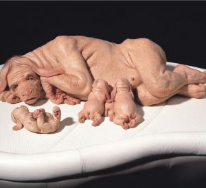
Chimera: a hybrid between stem cells of a dog and of a human
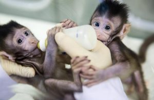
The first hybrid of an ape and a human-created in China